Table of Contents >> Show >> Hide
- When Doctors Start Looking for Esophageal Cancer
- The Core Test: Upper Endoscopy (EGD) With Biopsy
- Pathology: What the Lab Report Can Tell You
- Imaging and Procedures That Help Stage the Cancer
- Other Tests Used in Specific Situations
- How the Tests Fit Together: A Typical Diagnostic Path
- Questions to Ask Your Care Team
- Real-World Experiences: What These Tests Often Feel Like (and How People Cope)
- 1) The emotional roller coaster before the first big test
- 2) Endoscopy: “I blinked and it was over” (for many people)
- 3) Barium swallow: the chalky celebrity cameo
- 4) CT scan with contrast: warm sensation, fast results
- 5) PET/CT: the “quiet time” challenge
- 6) EUS and needle sampling: similar vibe to endoscopy, with extra information
- 7) The “results conversation”: where questions matter most
- Bottom Line
If swallowing has started to feel like your esophagus is auditioning to be a traffic jam, you’re not alone in wondering what’s going on.
Esophageal cancer is not the most common explanation for symptoms like trouble swallowingbut because it can be serious, doctors move
quickly and use very specific tests to get clear answers.
This guide walks through the main tests used to diagnose esophageal cancer (and the “supporting cast” that helps stage it). You’ll learn what
each test does, why it’s ordered, what results can mean, and what the experience is typically likewithout turning this into a medical thriller.
(Spoiler: the hero is usually an endoscope.)
When Doctors Start Looking for Esophageal Cancer
Testing usually begins when symptoms or risk factors suggest the esophagus needs a closer look. Many conditions can mimic each otheracid reflux,
inflammation, scarring (strictures), ulcers, or motility problemsso the goal is to separate “treatable and common” from “needs urgent attention.”
Symptoms that often trigger a workup
- Difficulty swallowing (dysphagia), especially if it’s getting worse over weeks to months
- Food “sticking” in the chest or throat, or frequent choking/coughing with meals
- Unexplained weight loss or reduced appetite
- Persistent chest discomfort or pain with swallowing
- Long-standing reflux, especially with new or worsening symptoms
- Hoarseness or chronic cough (in some cases)
Risk factors that may raise suspicion
Doctors also consider personal risk factors. These don’t diagnose anything on their own, but they help decide how urgently to test and what
tests to choose.
- Barrett’s esophagus (a change in the esophageal lining often linked to chronic GERD)
- Smoking and heavy alcohol use (more strongly linked with squamous cell carcinoma)
- Obesity and long-term reflux (linked with adenocarcinoma)
- Prior caustic injury to the esophagus or certain rare conditions affecting swallowing
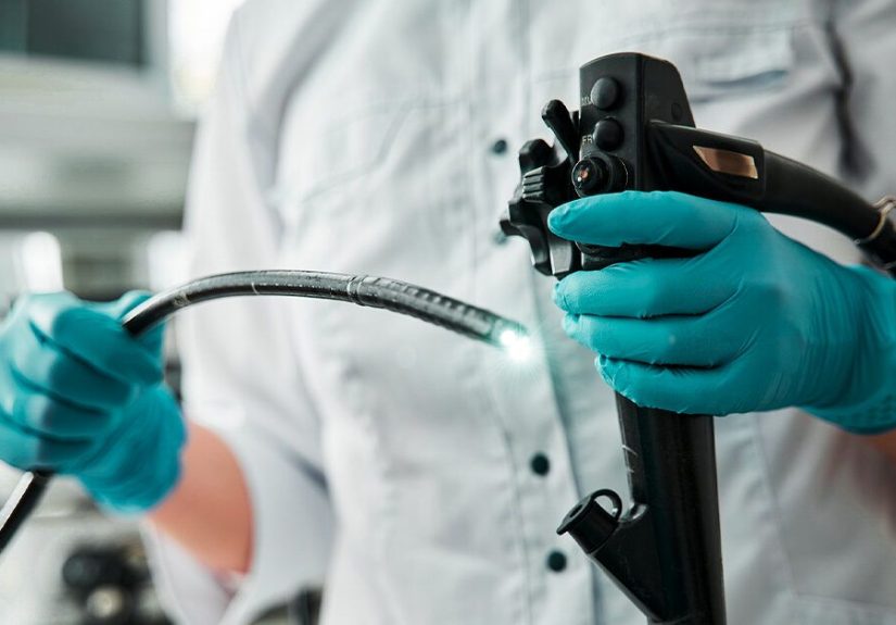
The Core Test: Upper Endoscopy (EGD) With Biopsy
If there’s one test to remember, it’s this: an upper endoscopy (EGD) with biopsy is the key test used to diagnose esophageal cancer.
Imaging can suggest a problem, but a biopsy is what confirms cancer.
What an EGD actually is
EGD stands for esophagogastroduodenoscopy. A gastroenterologist uses a thin, flexible tube with a camera and light to look directly
at the lining of the esophagus (and usually the stomach and first part of the small intestine). This is how doctors can spot:
- Suspicious masses or narrowed areas
- Ulcers or abnormal-looking tissue
- Barrett’s esophagus or pre-cancerous changes
Why the biopsy matters
During the endoscopy, the clinician can take tiny tissue samples (biopsies) using tools passed through the scope. A pathologist examines
these under a microscope to determine whether cancer is present and what type it is.
This is the medical version of “pics or it didn’t happen”except the “pics” are cells on a slide, and the “it” is a diagnosis that guides treatment.
What the patient experience is usually like
- Preparation: Often fasting for several hours beforehand. Your team will give specific instructions.
- During the test: Many people receive sedation. Most don’t remember much (or anything) about the procedure.
- Afterward: You’ll recover briefly, may feel groggy, and typically need someone else to drive you home if sedation was used.
Enhanced endoscopy techniques
Sometimes the endoscopy uses special imaging to better detect subtle abnormalitiesparticularly for Barrett’s esophagus surveillance or to define the
edges of suspicious tissue. Examples include dye-based techniques (chromoendoscopy) or “virtual” enhancements (technology that highlights mucosal patterns).
Pathology: What the Lab Report Can Tell You
Once the biopsy is taken, pathology becomes the decision-maker. The report doesn’t just say “cancer” or “not cancer.” It often answers several
practical questions that affect next steps.
Type of esophageal cancer
The two most common types are:
- Adenocarcinoma (often develops in the lower esophagus, commonly associated with Barrett’s esophagus and chronic reflux)
- Squamous cell carcinoma (more often in the upper or mid esophagus; historically associated with tobacco and alcohol exposure)
Grade and other features
The lab may describe how abnormal the cancer cells look compared with typical cells (often called grade), plus other details that can help
estimate how the tumor may behave.
Biomarker testing (often for treatment planning)
In many modern cancer centers, biopsy tissue may also be tested for biomarkers that help guide therapy decisionsespecially when advanced disease is suspected.
Examples can include HER2, PD-L1, MSI/MMR, and others depending on the situation.
These tests are typically not what “diagnoses” esophageal cancer, but they can shape the treatment plan once cancer is confirmed.
Imaging and Procedures That Help Stage the Cancer
After cancer is confirmed by biopsy, the next question is: How far has it grown or spread? This is called staging, and it’s
crucial for choosing treatment. Many tests below are ordered primarily for staging, but they are often described as part of the diagnostic process because
they help complete the picture.
Barium swallow (esophagram)
This test uses X-rays after you swallow a chalky contrast liquid called barium. The barium coats the lining of the esophagus so narrowing or irregular
contours show up more clearly.
- What it’s good for: Showing a stricture, mass effect, or abnormal swallowing mechanics
- Limitations: It cannot confirm cancer; it can point to a problem that needs endoscopy and biopsy
- Experience note: “Chalky milkshake” is a common descriptionno sprinkles, unfortunately.
CT scan (computed tomography)
CT scans of the chest and abdomen (often including the pelvis) look for spread to lymph nodes or nearby organs and provide a broader map of what’s going on.
CT is a workhorse for staging many cancers.
- What it’s good for: Lymph node enlargement, organ involvement, overall anatomy for planning
- Often includes contrast: You may drink contrast and/or receive IV contrast to improve visibility
PET scan (often PET/CT)
A PET scan uses a small amount of radioactive glucose (sugar) to highlight areas of higher metabolic activity. Many cancers “light up” because they use
more energy than surrounding tissue. PET is often combined with CT to match activity with anatomy.
- What it’s good for: Detecting spread that might not be obvious on CT alone
- Limitations: Not every abnormal “hot spot” is cancer (inflammation can also increase uptake), so results are interpreted carefully
- Experience note: The hardest part is usually the quiet resting period while the tracer distributesbring your best “still statue” skills.
Endoscopic ultrasound (EUS) with or without needle sampling
EUS combines endoscopy with ultrasound to see the esophageal wall layers and nearby lymph nodes in high detail. This helps evaluate how deep the tumor
has grown and whether nearby lymph nodes look suspicious.
- What it’s good for: Assessing depth of invasion (T stage) and nearby lymph nodes (N stage)
- Fine-needle aspiration (FNA): If a lymph node looks suspicious, a thin needle can sample it through the scope for lab analysis
- Why it matters: EUS can influence whether surgery, chemotherapy, radiation, or combined approaches are recommended
MRI (magnetic resonance imaging)
MRI is not always routine for esophageal cancer, but it can be used in specific situationssuch as clarifying findings on other scans or evaluating certain
tissues in more detail when needed.
Other Tests Used in Specific Situations
Not everyone needs every test. Doctors pick based on symptoms, tumor location, and what has already been learned. Think of it as choosing the right tools,
not collecting the whole hardware aisle.
Bronchoscopy
If a tumor is in the upper or mid esophagusor if imaging suggests possible involvement near the airwaydoctors may use bronchoscopy to look inside the
windpipe and major airways.
Laparoscopy or thoracoscopy (select staging procedures)
In certain cases, minimally invasive surgical procedures are used to look for small areas of spread that might not show up on scans. This can help avoid
major surgery when cancer has already traveled beyond where surgery would help.
Blood tests
Blood tests generally do not diagnose esophageal cancer, but they can reveal issues that matter for safety and planningsuch as anemia,
nutrition-related concerns, liver function, and overall fitness for treatment.
How the Tests Fit Together: A Typical Diagnostic Path
While every patient’s situation is unique, many workups follow a predictable pattern: confirm the diagnosis with a biopsy, then stage with imaging to guide
treatment. Here’s a realistic example flow.
Example timeline (one common pathway)
- Primary care or GI visit: Symptoms reviewed; risk factors assessed; referral for endoscopy
- EGD + biopsy: Suspicious area found and sampled
- Pathology confirms cancer: Type identified (adenocarcinoma vs squamous), basic tumor features described
- Staging begins: CT chest/abdomen/pelvis and often PET/CT
- EUS: Depth and lymph nodes evaluated; possible needle sampling if needed
- Special tests if indicated: Bronchoscopy or laparoscopy in select cases
- Multidisciplinary planning: Oncology, surgery, radiation oncology, GI, nutrition, and sometimes speech/swallow specialists coordinate
Quick “what each test is trying to answer” table
| Test | Main purpose | What it can (and can’t) confirm |
|---|---|---|
| Upper endoscopy (EGD) | Directly view lining; take biopsies | Can confirm cancer via biopsy; cannot fully stage spread by itself |
| Biopsy & pathology | Microscopic confirmation | Confirms cancer type and features |
| Barium swallow | Outline narrowing/irregularity | Suggests abnormalities; cannot confirm cancer |
| CT scan | Check lymph nodes/organs | Estimates spread; may miss tiny areas |
| PET/CT | Look for metabolically active spread | Helps detect hidden metastases; not every “hot spot” is cancer |
| EUS (± FNA) | Depth of invasion and nearby nodes | Improves local staging; can sample suspicious nodes |
Questions to Ask Your Care Team
Being tested can feel like you’re living inside a calendar invite. These questions can help you get clear, practical answers:
- Which test will confirm the diagnosis, and when will results be ready?
- Are you testing for staging (how far it has spread) or diagnosis (confirming cancer), or both?
- Will I have sedation for the endoscopy or EUS? What should I expect afterward?
- Which scans are recommended for my tumor location, and why?
- Will the biopsy sample be tested for biomarkers that could affect treatment choices?
- Should I meet with a nutrition or swallowing specialist now?
- What symptoms should make me call right away while waiting for tests?
Real-World Experiences: What These Tests Often Feel Like (and How People Cope)
Medical brochures can be a little… optimistic. (“You may experience mild discomfort.” Translation: “You might feel weird, and that’s normal.”)
Here are common experiences people report during esophageal cancer testingplus the small coping tricks that make a big difference.
1) The emotional roller coaster before the first big test
Many people say the hardest part is the waitingwaiting for an appointment, waiting for sedation to wear off, waiting for pathology.
It’s common to feel anxious, distracted, or stuck in “doom-scroll mode,” even if you’re usually calm under pressure.
A surprisingly helpful move is to write down your symptoms and questions ahead of time. In the moment, it’s easy to forget details like
“it’s worse with bread” or “it started after that reflux flare.” A short note on your phone can keep the visit focused and help you feel more in control.
2) Endoscopy: “I blinked and it was over” (for many people)
When sedation is used, a lot of patients describe EGD as the easiest test in hindsight: they remember checking in, maybe a few minutes in the procedure room,
then waking up in recovery wondering if anything happened yet. Afterward, some feel groggy, hungry, or mildly sore in the throat.
People often recommend bringing a support person (and a snack for after, if your team says it’s okay).
The most important practical tip: if you’re sedated, plan to take it easy the rest of the dayyour brain may be “online,” but it’s running the
free trial version.
3) Barium swallow: the chalky celebrity cameo
The barium swallow is typically quick and doesn’t involve needles or sedation, which many people appreciate. The most memorable part is the texture and taste:
“like a chalky smoothie” is a frequent review. During the test, you may be asked to change positions so X-rays can capture different angles while you swallow.
People often say it feels a bit awkward (like a photoshoot where the director keeps saying, “Turn… now swallow… hold it…”), but not painful.
Some also mention temporary constipation afterwardyour care team may recommend hydration or specific guidance if needed.
4) CT scan with contrast: warm sensation, fast results
CT scans are usually fast. If IV contrast is used, a common experience is a brief warm feeling or a metallic taste.
Most patients say the anticipation sounds worse than the reality. What helps: wear comfortable clothing, show up hydrated (unless told otherwise),
and tell the team if you’ve had reactions to contrast in the past.
5) PET/CT: the “quiet time” challenge
PET scans often include a waiting period after the tracer injection. People describe this as the most boring part: you may need to rest quietly, avoid
vigorous movement, and stay warm. The scan itself is typically painless. A tip many patients share: treat the wait like a mini meditation session.
Slow breathing, a calm playlist (if allowed), or a simple grounding exercise can make it feel less like “time is crawling” and more like “I’m doing something useful.”
6) EUS and needle sampling: similar vibe to endoscopy, with extra information
Patients often experience EUS much like EGDespecially if sedation is used. The difference is mostly behind the scenes:
the ultrasound images and possible lymph node sampling can provide key staging details. People frequently say that understanding
why EUS is being done helps them tolerate the stress of “one more procedure.” If you’re feeling overwhelmed, ask your team:
“What decision will this test help you make?” A clear answer can turn anxiety into purpose.
7) The “results conversation”: where questions matter most
Many people describe the first results visit as a blur. A practical strategy is to bring a trusted person to take notesor ask permission to record the
discussion on your phone for later review. Patients also say it helps to request the “plain English” version of the plan:
What stage do you think this is? What’s the goal of treatmentcure, control, or symptom relief? What are the next two steps on the calendar?
If you take one thing from these shared experiences, let it be this: you don’t need to be brave in a cinematic way.
You just need a clear plan, good communication, and support while the testing does its jobturning uncertainty into actionable information.
Bottom Line
Diagnosing esophageal cancer usually hinges on upper endoscopy with biopsy. After that, staging testssuch as CT, PET/CT, and EUShelp determine
how advanced the disease is and what treatment approach makes the most sense. Not every patient needs every test, and a good care team will explain
what each test is expected to change in the plan.
If you’re going through testing now, it’s okay to feel nervous. The purpose of this process is clarityso decisions can be made based on evidence, not fear.