Table of Contents >> Show >> Hide
- What Is Horner’s Syndrome?
- The “Classic” Signs (and Why They Happen)
- Why Horner’s Syndrome Matters: It’s Often a Clue, Not the Final Answer
- Causes: Where the “Wire” Can Get Interrupted
- Symptoms Checklist: What People Notice (and What They Miss)
- Diagnosis: How Clinicians Confirm Horner’s Syndrome
- Treatments: Fix the Cause, Not the Eyelid
- Outlook: What Living With Horner’s Syndrome Can Look Like
- When to Seek Urgent Care
- Questions to Ask at Your Appointment
- Real-World Experiences: What People Often Report (Composite Stories)
- Conclusion
Imagine waking up and discovering one eyelid has decided to take a personal day. Your pupil on that side looks smaller, your face feels a little “off,” and suddenly you’re Googling at Olympic speed. One possible explanation is Horner’s syndromea cluster of signs that can look subtle in the mirror but can be a big deal medically, depending on why it’s happening.
Horner’s syndrome isn’t a disease you “catch.” It’s more like a check-engine light for the sympathetic nerves that run from your brain to your eye and face. Sometimes the cause is harmless or already known (like recent surgery). Other times, it can signal something urgentlike a tear in an artery in the neck. The goal of this guide is to make the topic understandable, practical, and a little less scarywithout sugarcoating the parts that deserve fast attention.
What Is Horner’s Syndrome?
Horner’s syndrome happens when there’s a disruption in the sympathetic nerve pathway that helps control:
- How wide your pupil gets in dim light
- A small muscle that helps keep your upper eyelid lifted
- Sweating and certain blood vessel responses on the face
Because the sympathetic pathway is long (brain → spinal cord → chest/neck → eye), Horner’s syndrome can be caused by issues in multiple locations. That’s why clinicians treat it like a localization puzzle: the pattern of signs, plus your symptoms and timeline, helps point to where the nerve interruption might be.
The “Classic” Signs (and Why They Happen)
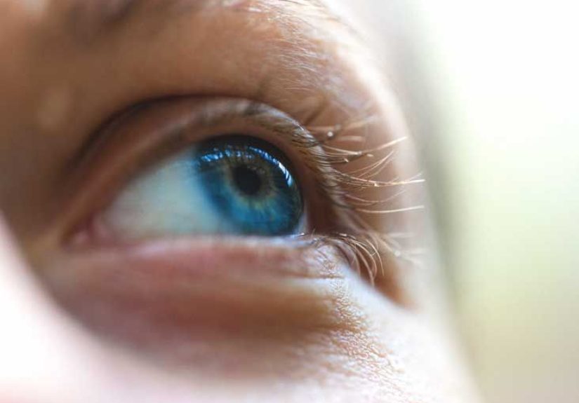
1) Miosis (a smaller pupil on one side)
The sympathetic system helps your pupil dilate (get bigger), especially in the dark. If that sympathetic input is reduced, the affected pupil tends to stay smaller. A key clue is that the difference between pupilscalled anisocoriais often more noticeable in dim lighting, because the normal pupil expands and the affected one lags behind.
2) Ptosis (a drooping upper eyelid)
Horner’s syndrome typically causes a mild droop, not the dramatic “eye is shutting” look people associate with certain nerve palsies. That’s because it affects a small sympathetic muscle (Müller’s muscle), not the main eyelid-lifting muscle.
3) Anhidrosis (reduced sweating on one side of the face)
Not everyone notices sweating changes, and the location matters. Depending on where the pathway is interrupted, sweating changes may involve the face, or sometimes be minimal. Think of it as the body’s way of saying, “My wiring isn’t getting the memo.”
Other possible clues
- “Sunken eye” appearance: often more of an optical illusion from the eyelid changes than a true sinking of the eye.
- Heterochromia (different iris color): more likely in congenital or long-standing cases that start in infancy.
Why Horner’s Syndrome Matters: It’s Often a Clue, Not the Final Answer
Here’s the part worth remembering: Horner’s syndrome itself usually doesn’t damage vision. The bigger concern is what caused the nerve disruption.
Sometimes the cause is obvious and non-emergencylike a known neck surgery, a benign mass already being monitored, or a long-standing congenital finding. But when Horner’s syndrome is new, especially in adults, clinicians often treat it as a sign that deserves prompt evaluation. One reason: a new Horner’s syndrome with head, face, or neck pain can be linked to a carotid artery dissection, which can raise stroke risk.
Causes: Where the “Wire” Can Get Interrupted
The sympathetic pathway involved in Horner’s syndrome is often described as a three-neuron chain. This isn’t just trivialocation helps guide which tests might be needed.
| Pathway level | General location | Examples of possible causes |
|---|---|---|
| First-order (central) | Brainstem/spinal cord | Stroke, tumor, demyelinating disease, spinal cord lesions |
| Second-order (preganglionic) | Chest/neck (sympathetic chain) | Lung apex tumors (Pancoast-type), chest/neck surgery, trauma, certain neck masses |
| Third-order (postganglionic) | Along carotid artery into the skull/eye | Carotid artery dissection, cavernous sinus issues, cluster headache-related autonomic symptoms |
Common themes across many causes
- Vascular issues: especially carotid artery dissection (a tear in the artery wall), which may come with neck or head pain.
- Masses or tumors: along the pathway (neck, chest, or brain). In children, certain tumors like neuroblastoma are a key consideration.
- Trauma or procedures: injuries or surgeries involving the neck, chest, or upper spine can affect the pathway.
- Inflammation/infection: less common, but infections or inflammatory processes in the neck/chest region can sometimes play a role.
Congenital and pediatric causes
In infants and children, Horner’s syndrome can be present from birth or appear later. Birth-related neck/shoulder trauma is a commonly discussed cause of congenital cases. When a child has new Horner’s syndrome without a clear explanation, clinicians may consider a workup that can include imaging and lab testing, because a small portion can be linked to serious underlying conditions.
Symptoms Checklist: What People Notice (and What They Miss)
Horner’s syndrome can be sneaky. Many people don’t feel “sick”they just notice something looks different. Common observations include:
- One pupil is consistently smaller
- The difference between pupils is more obvious in dim rooms or at night
- A mild eyelid droop on one side
- Less sweating or a different “flush” pattern on one side of the face (harder to spot)
- In children: an eyelid droop plus subtle iris color differences, especially if it started early
What Horner’s syndrome doesn’t usually cause by itself: severe vision loss, major eye pain, or complete eyelid shutdown. If those are present, clinicians think about other diagnoses too.
Diagnosis: How Clinicians Confirm Horner’s Syndrome
Diagnosis typically starts with a careful history and eye examespecially looking at pupil size in bright and dim light. Then, depending on the situation, clinicians may use pharmacologic testing and/or imaging.
The eye exam: light vs. dark matters
A classic pattern is anisocoria that’s greater in the dark, because the affected pupil doesn’t dilate normally. Clinicians also look for mild ptosis and other neurologic signs that could suggest where along the pathway the problem might be.
Apraclonidine testing (and what “reversal” means)
A commonly used confirmation test involves eye drops such as apraclonidine. In many cases of Horner’s syndrome, the affected eye develops “denervation hypersensitivity,” meaning it overreacts to certain stimulants. After drops, the smaller pupil may dilate more than expectedsometimes causing a reversal of anisocoria (the previously smaller pupil becomes similar to or larger than the other).
Important nuance: very early Horner’s syndrome may not always show a dramatic drop-test response, and clinicians interpret results in context rather than treating one test as an oracle.
Imaging: the “find the cause” phase
Once Horner’s syndrome is suspected or confirmed, the next question is: Why is it happening? Imaging choices depend on the clinical picture, but can include:
- CTA or MRA of the head and neck if a carotid artery problem is a concern (especially with pain)
- MRI of the brain and neck if central causes are possible
- Chest imaging when a lesion in the upper chest is suspected
Testing in children (including neuroblastoma considerations)
In pediatric cases without a clear explanation, clinicians may consider a combination of imaging and urine testing for catecholamine metabolites. However, urine tests aren’t perfectsome children with neuroblastoma may not have elevated results at presentationso imaging and clinical judgment matter a lot.
Treatments: Fix the Cause, Not the Eyelid
There’s no single “Horner’s syndrome pill.” Treatment is typically about identifying and addressing the underlying cause. When that cause is treated, Horner’s signs may improve, and sometimes they resolve.
Examples of how treatment depends on the cause
- Carotid artery dissection: usually needs urgent medical evaluation and management to reduce stroke risk.
- Cluster headaches: treating the headache disorder may lessen associated autonomic symptoms, including Horner-like findings in some cases.
- Tumors or masses: treatment depends on type and location (surgery, oncology care, or other targeted therapies).
- Post-surgical or post-procedure cases: sometimes improve with time; follow-up focuses on recovery and making sure nothing else is going on.
Can symptoms be treated directly?
Because ptosis in Horner’s syndrome is usually mild, many people don’t need cosmetic treatment. In select situations, an ophthalmologist may discuss options if droop is persistent and bothersome. Some clinicians note that apraclonidine can also temporarily raise the eyelid a bituseful diagnostically and occasionally cosmeticallybut it’s not a DIY beauty hack and should be used only under medical guidance.
Outlook: What Living With Horner’s Syndrome Can Look Like
Horner’s syndrome is often more alarming-looking than dangerous on its own. The long-term outlook depends mainly on the underlying cause and how quickly it’s addressed.
- Sometimes the signs improve after treating the cause.
- Sometimes the signs persist even after the cause is treatedespecially if the nerve pathway was permanently injured.
- Vision is typically okay, though people may notice cosmetic asymmetry or light sensitivity differences.
When to Seek Urgent Care
If Horner’s syndrome might be new, it’s worth medical evaluation. And in some cases, it’s worth urgent evaluation. Seek emergency care if you notice a new droopy eyelid or new unequal pupils plus any of the following:
- New head, face, or neck pain
- Weakness, numbness, trouble speaking, severe dizziness, or other stroke-like symptoms
- Recent significant trauma
For children, prompt evaluation is important when Horner’s signs are new or unexplainedespecially if there are additional concerning symptoms (like a new neck mass, unexplained weight loss, or persistent fatigue).
Questions to Ask at Your Appointment
- Do my symptoms fit Horner’s syndrome, or something else?
- Is this likely new or long-standing?
- Do I need imaging right away? If so, what kind and why?
- Are there red flags that mean I should go to the ER?
- If a cause is found, what’s the treatment plan and follow-up schedule?
Real-World Experiences: What People Often Report (Composite Stories)
Note: The experiences below are composites based on common clinical scenarios and patient-reported themes. They’re meant to help you recognize patterns and prepare questionsnot replace medical care.
The “I thought it was just tired eyes” moment
A surprisingly common story starts with a mirror, a selfie, or a friend saying, “Hey… is your eyelid drooping?” The person feels finemaybe a little stressed, a little sleep-deprived, nothing dramatic. They assume allergies, aging, or too much screen time. Then they notice the pupil on that side looks smaller, especially at night. That “wait, what?” moment is often what pushes someone to see an eye doctor or urgent care.
Emotionally, this stage can be weirdly frustrating: the symptoms look obvious to the person, but can be subtle under bright clinic lights. People often describe relief when a clinician takes photos in dim lighting or measures the pupils carefully, because it turns a vague worry into a real, nameable finding.
The “this came with pain, and that changed everything” scenario
Another pattern is Horner’s signs paired with head, face, or neck pain. People describe it as sharp, persistent, or just “not my normal headache.” This is one of the most anxiety-provoking situationsand for good reasonbecause clinicians may worry about vascular causes such as carotid artery dissection. In these cases, the experience is often fast-paced: exam, urgent imaging, a lot of new vocabulary, and a strong message not to ignore neurologic symptoms.
Many people say the most helpful thing in hindsight was having a clear explanation of why doctors moved quickly. Even when the final results are reassuring, understanding the logic (“we have to rule out the dangerous stuff first”) can make the process feel less like a medical roller coaster.
Parents noticing changes in a baby or toddler
For parents, the story can be even more unsettling: a baby’s eyelid looks droopy in certain photos, or one pupil seems smaller. Sometimes the pediatrician notices it during a routine visit. Families often describe a tug-of-war between “It’s probably nothing” and “But what if it’s something?” Pediatric evaluations can involve imaging and sometimes urine testing, which can be stressful even when the child seems otherwise fine.
Parents frequently say the emotional challenge isn’t just the testingit’s the waiting and uncertainty. Having a clinician explain what they’re looking for (and what makes serious causes less likely) can be calming. If Horner’s is congenital or linked to a known birth-related injury, families often shift from fear to logistics: tracking changes, monitoring development, and deciding whether the eyelid droop is noticeable enough to consider specialty care later.
Living with a persistent, mild asymmetry
Some people end up with a long-term, mild ptosis and small pupil difference even after the underlying issue is treatedor after a workup finds no dangerous cause. The practical impact can be surprisingly… normal. People often report that they notice it more than anyone else does, but they may still feel self-conscious on camera or under certain lighting.
What helps? Small wins: good lighting for video calls, understanding that mild asymmetry is common in humans anyway, and having a clinician validate the concern. For those with persistent droop that affects confidence or function, a conversation with an ophthalmologist about options can feel empoweringnot because it’s “necessary,” but because it puts the person back in the driver’s seat.
Conclusion
Horner’s syndrome is a patterntypically a smaller pupil, mild eyelid droop, and sometimes reduced facial sweatingthat points to a disruption in the sympathetic nerves to the eye and face. The most important step isn’t “treating Horner’s” so much as finding out why it’s happening. When it’s newespecially if it’s paired with head or neck painit deserves prompt medical evaluation. The good news is that many causes are treatable, and the eye findings themselves are often manageable once the underlying issue is addressed.