Table of Contents >> Show >> Hide
- What Is a Testicular (Scrotal) Ultrasound?
- Why Would You Need One?
- What Is Doppler, and Why Does Everyone Get Excited About It?
- How to Prepare (Spoiler: It’s Usually Easy)
- Step-by-Step: What Happens During the Procedure?
- Risks and Safety
- What Can the Results Show?
- What Happens After the Ultrasound?
- Limitations: What Ultrasound Can’t Do
- When to Seek Care Immediately
- FAQ: The Questions People Actually Ask
- Real-World Experiences (500+ Words): What It’s Like, Emotionally and Physically
- Conclusion
Let’s talk about a testicular ultrasoundalso called a scrotal ultrasound.
If your brain immediately goes, “Cool… so I’m bringing my anxiety and my dignity to the appointment,” you’re not alone.
The good news: this test is noninvasive, uses sound waves (not radiation), and usually takes
about 15–30 minutes. It’s one of the fastest, most helpful ways to figure out what’s going on
when something in the scrotum feels offpain, swelling, a lump, or “this looks different than yesterday.”
In this guide, you’ll learn what a testicular ultrasound checks, why Doppler matters (yes, it’s a real word),
what the procedure feels like, what results can mean, and how to use the information without spiraling into
late-night internet doom-scrolling.
What Is a Testicular (Scrotal) Ultrasound?
A testicular ultrasound is an imaging test that creates pictures of the testicles and nearby structures
inside the scrotum. The scan uses high-frequency sound waves to produce images on a screen.
You’ll also hear it called testicular sonogram or ultrasound of the scrotum.
Ultrasound is especially useful because it can help distinguish what’s happening inside the testicle
versus around it, and it can evaluate soft tissues and fluid collections that don’t show up well on some other tests.
Why Would You Need One?
Doctors order scrotal ultrasound for lots of reasons, but the big categories are:
pain, swelling, a lump/mass, injury,
or questions about blood flow and fertility.
Common Reasons (Indications)
- Sudden scrotal pain (especially if severe or one-sided)
- Swelling of a testicle or the scrotum
- A lump you or your clinician can feel
- Infection or inflammation (epididymitis/orchitis)
- Testicular torsion (twisting that can cut off blood flowan emergency)
- Varicocele (enlarged veinssometimes linked with fertility issues)
- Hydrocele (fluid around the testicle)
- Trauma (sports injury, accident, direct hit)
- Infertility evaluation (when scrotal anatomy might be contributing)
Real-world example: you notice a new lump after a shower. Your clinician may order a testicular ultrasound to help
determine if it looks like a fluid-filled cyst, inflammation, or a solid mass that needs urgent follow-up.
Importantly, not all testicular lumps are cancerespecially smaller findingsso imaging helps guide the next step.
What Is Doppler, and Why Does Everyone Get Excited About It?
Many scrotal ultrasounds include Doppler ultrasound, which evaluates blood flow.
On the screen, Doppler can show flow patterns (often with color) in the testicles and surrounding vessels.
This matters most when your doctor is worried about conditions where circulation changes quickly:
testicular torsion (reduced/absent flow) versus infection/inflammation (often increased flow).
In acute scrotal pain, Doppler is commonly the first-line imaging choice because it can help separate
“needs surgery now” from “needs antibiotics and rest,” though clinical judgment still rules the day.
How to Prepare (Spoiler: It’s Usually Easy)
For most people, there’s little to no special preparation.
You may be asked to undress from the waist down and wear a gown. It can help to wear comfortable underwear
and clothing that’s easy to change in and out of. If you’re in pain, it’s okay to mention that right away
so the staff can position you as comfortably as possible.
Helpful Prep Tips
- Shower normally (no special shaving requirementsyour body, your rules).
- If you’re in significant pain, consider bringing supportive underwear for after the exam.
- Bring a list of symptoms: when it started, what makes it worse/better, fever, nausea, recent injury, etc.
- If this is for a lump, note whether it changes with position or time of day.
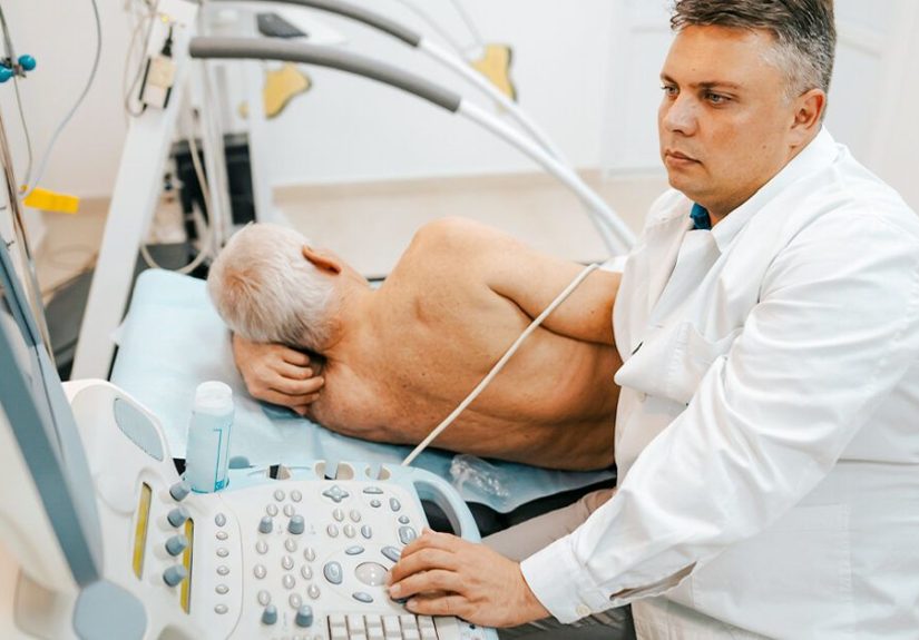
Step-by-Step: What Happens During the Procedure?
The procedure is typically done by a sonographer (ultrasound technologist) and interpreted by a radiologist.
Your clinician then reviews the results with you.
-
Positioning: You’ll lie on your back. The scrotum is often supported with a towel to keep things steady.
The penis is typically moved out of the way and covered for modesty while the scrotum is examined. -
Gel time: A water-based gel is placed on the skin. It may feel cool at first.
(Yes, that moment is startling. No, you’re not the first person to flinch.) -
Scanning: The technologist gently moves a handheld probe (transducer) over the scrotum.
They’ll take images from different angles, sometimes comparing both sides. -
Doppler evaluation: If Doppler is included, they’ll assess blood flow patterns to help evaluate
torsion, inflammation, varicocele, and other vascular changes. -
Finish and clean-up: The gel is wiped off, and you can get dressed and go about your day.
No recovery time is typically needed.
How Long Does It Take?
Most scrotal ultrasounds are completed in about 15 to 30 minutes, though the timing can vary if the case is complex.
Does It Hurt?
The exam itself is usually not painful, but if you already have tenderness or swelling,
you may feel discomfort when the probe moves over sensitive areas. If it hurts, say sotechnologists can adjust
pressure and positioning.
Risks and Safety
Testicular ultrasound is considered very safe. It does not use ionizing radiation.
For typical scrotal ultrasound, there are no known risks. The main “risk” is mild discomfort if the area is already painful,
plus a temporary awkwardness level that spikes for 20 minutes and then disappears forever.
What Can the Results Show?
Ultrasound results usually describe the appearance of the testicles and surrounding structures:
size, texture, presence of fluid, masses, inflammation, and blood flow patterns (if Doppler was used).
Your report may include terms like “cystic,” “solid,” “hypoechoic,” or “vascularity.”
Translation: it’s describing what the images look like and how blood flow behavesclues to the diagnosis.
Examples of Findings (and What They Often Suggest)
- Hydrocele: Fluid around the testicle. Often benign; may be related to irritation, injury, or infection.
- Varicocele: Enlarged veins (often on the left). Can be associated with aching discomfort or fertility concerns.
-
Epididymitis/orchitis: Signs of inflammation; Doppler may show increased blood flow.
Treatment often involves medications and supportive care. -
Testicular torsion: Concerning for decreased/absent blood flow and twisting features.
This is a time-sensitive emergency that can require urgent surgical evaluation. -
Mass inside the testicle: Requires prompt evaluation by urology. Many intratesticular solid masses are treated as suspicious
until proven otherwise, but benign causes existimaging is part of the workup, not the final verdict. - Cyst or benign scrotal lesion: Often a non-cancerous, fluid-filled finding outside the testicle.
How Fast Will You Get Results?
Many facilities can provide results relatively quickly, but timing depends on the setting.
In an emergency department, results may be reviewed urgently. In outpatient imaging, you might get a report
within a day or two, then your clinician follows up with the plan.
What Happens After the Ultrasound?
Most people return to normal activities immediately. The important part is what your clinician recommends next.
That might include:
- Medication (for infection/inflammation)
- Follow-up imaging (if something needs monitoring)
- Referral to a urologist (especially for masses, persistent symptoms, fertility issues)
- Urgent surgical evaluation (if torsion or serious trauma is suspected)
Limitations: What Ultrasound Can’t Do
Ultrasound is powerful, but it’s not a magical lie detector for every scrotal mystery.
A few limitations to keep in mind:
- It’s operator- and interpretation-dependent: image quality and accuracy rely on technique and experience.
- It doesn’t replace clinical judgment: severe symptoms still need urgent evaluation even if images are “not dramatic.”
- Some findings are nonspecific: a report may say “indeterminate,” meaning it doesn’t scream one diagnosis.
-
Cancer vs. benign: ultrasound can strongly suggest features, but diagnosis may still require specialist evaluation,
labs, or surgery/biopsy decisions in selected cases.
When to Seek Care Immediately
Call emergency services or seek urgent care if you have:
- Sudden, severe testicular/scrotal pain
- Swelling with nausea/vomiting
- Fever with significant scrotal pain
- Severe pain after trauma
- A testicle that looks higher than usual or sits at a new angle
These symptoms can be seen in serious conditions like torsion or complicated infections where timing matters.
FAQ: The Questions People Actually Ask
Will the ultrasound expose me to radiation?
No. Ultrasound uses sound waves and does not involve ionizing radiation.
Is it awkward?
It can feel awkward because of the body part involved, but medically it’s routine.
The staff does this all day, every day, and their main goal is accurate imaging and your comfort.
If you want a chaperone or feel anxious, you can askseriously.
Can I drive myself home?
Typically yes. There’s no sedation and no expected downtime.
If you’re in intense pain or being evaluated for emergency conditions, follow the clinician’s guidance.
Does a normal ultrasound mean I’m “in the clear”?
A normal result is reassuring, but it’s not a universal hall pass.
If symptoms persist or worsen, follow upbecause bodies love changing the plot when you least expect it.
Real-World Experiences (500+ Words): What It’s Like, Emotionally and Physically
People rarely schedule a testicular ultrasound because they’re bored on a Tuesday. Usually, it starts with a moment:
a sharp pain during a workout, a dull ache that won’t quit, swelling that shows up out of nowhere, or a new lump
discovered in the shower. The first experience most people have is not the scan itselfit’s the mental soundtrack:
“Is this serious?” “Did I wait too long?” “Do I tell my partner?” “Am I overreacting?” (For the record: if you noticed a change,
you’re doing the responsible thing by getting it checked.)
The day of the ultrasound, the most common surprise is how normal and procedural it feels. You check in, answer a few questions,
change into a gown, and then it’s mostly quiet concentration while the technologist captures images.
The gel can feel cold, and the probe pressure can be uncomfortable if you’re already tenderespecially with swelling or inflammation.
Many people describe the sensation as “weird but tolerable,” like someone gently pressing a spoon onto a bruise.
If pain is part of why you’re there, it’s completely reasonable to say, “That spot is really sore,” so they can adjust.
Emotionally, a lot of people feel exposednot just physically, but psychologically. There’s vulnerability in needing help for something
tied to masculinity, fertility, or body image. Some patients report feeling embarrassed walking into imaging, then realizing the staff is
calm, professional, and absolutely not judging. Others feel relief during the scan because it’s action: you’re no longer guessing.
You’re collecting real information. That shiftfrom uncertainty to datacan be a big deal.
Waiting for results is often the toughest part. Even when a clinic is fast, the brain can fill in gaps with worst-case scenarios.
A common experience is learning that the scary thing you felt is a benign issuelike a hydrocele, a cyst, or a varicocele.
People often describe a “release valve” moment: shoulders drop, breathing slows, and they realize how tense they’ve been.
On the flip side, if an ultrasound finds something that needs follow-up, patients often feel a mix of fear and gratitude:
fear because it’s unknown, gratitude because it was caught early and now there’s a plan.
Many men also mention that the appointment becomes a turning point for self-awareness. Afterward, they’re more likely to notice changes,
do a quick self-check occasionally, and seek care sooner rather than later. Some share that they wish they’d come in earlier, not because
something terrible happened, but because the anxiety of “not knowing” was heavier than the test itself.
A testicular ultrasound can’t solve every problem instantly, but it can transform a vague worry into a clear next stepwhether that’s
reassurance, medication, monitoring, or specialist care.
If you’re reading this because you’re worried: the most common “experience” people report in hindsight is that the scan was easier than
they imagined. The hardest part was the lead-up. The most helpful part was clarity.
Conclusion
A testicular ultrasound is a quick, safe, and highly informative test used to evaluate scrotal pain, swelling, lumps, trauma,
and blood-flow concerns. With Doppler imaging, it can provide crucial clues in urgent scenarios like suspected torsion, while also helping
identify common conditions such as varicoceles, hydroceles, and infections. If you’ve noticed a change, getting checked is the grown-up move.
And if the exam feels awkward? Congratulationsyou are a human with a nervous system. You’ll get through it.