Table of Contents >> Show >> Hide
- What Is a Salivary Mucocele in Dogs?
- The 12-Step Guide
- Step 1: Notice the shape, feel, and location of the swelling
- Step 2: Do not assume “painless” means harmless
- Step 3: Watch for eating and drinking changes
- Step 4: Treat breathing trouble as urgent
- Step 5: Check for related clues, not just the lump itself
- Step 6: Resist the urge to poke, squeeze, or drain it at home
- Step 7: Schedule a veterinary exam and be ready to describe the timeline
- Step 8: Expect fine-needle aspiration, and possibly imaging
- Step 9: Understand what else it could be
- Step 10: Learn the main treatment options
- Step 11: Prepare for recovery like an organized grown-up who now owns a cone
- Step 12: Know what prognosis and recurrence usually look like
- Why Early Treatment Matters
- Common Owner Experiences: What This Usually Looks Like in Real Life
- Final Takeaway
If you discover a squishy lump under your dog’s jaw, your brain may sprint straight to the worst-case scenario. Fair enough. Dog owners are not known for seeing a mystery mass and calmly saying, “Ah yes, this is probably a manageable salivary issue.” But in some dogs, that strange swelling turns out to be a salivary mucocele, also called a sialocelea condition where saliva leaks out of a damaged salivary gland or duct and collects in nearby tissue.
The good news? A salivary mucocele in dogs often looks dramatic before it acts dramatic. Many dogs stay bright, hungry, and deeply committed to snack negotiations. The not-so-good news is that this problem usually does not fix itself for long. It can keep recurring, grow larger, and in certain locations make swallowing or breathing harder than it should be. That is why early recognition matters.
This guide walks you through exactly how to recognize a salivary mucocele in dogs, what symptoms deserve urgent attention, how vets diagnose it, and what treatment and recovery usually look like. To keep things practical, the article is organized into 12 clear steps, so you can go from “What on earth is this lump?” to “Okay, I know what to do next.”
What Is a Salivary Mucocele in Dogs?
A salivary mucocele is a pocket of leaked saliva that forms when a salivary gland or salivary duct is injured, blocked, or ruptured. Instead of flowing into the mouth where it belongs, saliva seeps into surrounding tissue and the body walls it off with inflamed connective tissue. That means a mucocele is not a classic “cyst” in the neat little balloon sense. It is more like the body building a messy containment zone around escaped saliva.
Dogs have several major salivary glands, but the mandibular and sublingual glands are involved most often. Depending on where the saliva collects, a mucocele may show up as:
- Cervical mucocele: swelling in the upper neck or under the jaw
- Sublingual mucocele: a ranula under or beside the tongue
- Pharyngeal mucocele: swelling in the throat
- Zygomatic mucocele: swelling near the eye
Now let’s get into the 12-step process of spotting it and dealing with it the smart way.
The 12-Step Guide
Step 1: Notice the shape, feel, and location of the swelling
The first clue is usually a soft lump. Most owners find it under the lower jaw, along the upper neck, or on one side of the throat area. It often feels squishy or fluid-filled rather than rock-hard. Some dogs act completely normal, which can make the lump even more confusing. A mucocele may be large, but it is often not painful when you touch it.
If the swelling is near the eye, under the tongue, or in the throat, the appearance may be less obvious. In those cases, the first thing you notice may be a change in chewing, swallowing, or facial symmetry rather than a classic neck lump.
Step 2: Do not assume “painless” means harmless
This is where dog health likes to play tricks. A dog with a salivary mucocele may still wag, eat, beg, and patrol the kitchen like a furry hall monitor. Because many mucoceles are nonpainful at first, owners may delay care. But saliva keeps leaking, inflammation keeps building, and the swelling can continue to expand.
So yes, your dog may look completely unbothered. Your dog may also be wrong. A slowly growing lump still deserves veterinary attention, even when your pet is acting like the mayor of a very normal Tuesday.
Step 3: Watch for eating and drinking changes
When a salivary mucocele forms under the tongue or near the throat, it can interfere with normal eating. Dogs may chew oddly, drop food, eat more slowly, turn their head while chewing, or seem reluctant to tackle dry kibble. Some will swallow repeatedly, drool more, or avoid hard treats that used to disappear in three crunches flat.
A ranula, which is a sublingual mucocele in dogs, may also get bumped by the teeth during chewing and can bleed. If you notice blood-tinged saliva, oral swelling, or a fleshy bubble under the tongue, that moves this out of the “let’s just monitor it” category.
Step 4: Treat breathing trouble as urgent
A pharyngeal salivary mucocele can press into the throat and narrow the airway. That can lead to noisy breathing, gagging, coughing, stridor, open-mouth breathing, or visible distress. Dogs with throat-based mucoceles may also have trouble swallowing because there simply is not enough space where there ought to be space.
If your dog seems short of breath, cannot swallow normally, or appears panicked while trying to breathe, get urgent veterinary care right away. This is not the moment for internet deep-dives, group chats, or a heartfelt promise to “see how she does by morning.”
Step 5: Check for related clues, not just the lump itself
Salivary gland swelling in dogs can come with extra signs that help point to the location of the problem. Look for:
- Excess drooling
- Blood in the saliva
- Reluctance to chew
- Bad breath from oral irritation
- Bulging of one eye if the zygomatic gland is involved
- Swallowing difficulty or repeated gagging
These clues matter because a neck mucocele behaves differently from a ranula or a throat mucocele. The exact location helps shape the diagnostic plan and the surgical approach.
Step 6: Resist the urge to poke, squeeze, or drain it at home
This one deserves bold emotional emphasis: do not try to drain a dog’s salivary mucocele yourself. Even veterinarians know that simple drainage is usually temporary. The pocket often refills because the source of the leak is still there. Repeated poking can also introduce bacteria and make a frustrating problem worse.
Trying to “pop” it at home can cause pain, contamination, bleeding, and a fast track to an infection you definitely did not order. In other words, this is not a pimple, not a cyst video, and absolutely not a DIY weekend project.
Step 7: Schedule a veterinary exam and be ready to describe the timeline
Your veterinarian will want details: when you first noticed the swelling, whether it changes size, whether your dog has trouble eating or breathing, and whether there was any recent trauma, bite wound, chewing on sticks, rough collar pull, or facial surgery. In many dogs, the exact cause is never identified, but the history still helps narrow the possibilities.
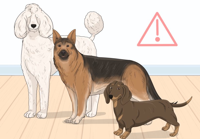
Some breeds appear more commonly represented in case reports and specialty practices, including Dachshunds, Poodles, and German Shepherd Dogs. Still, a salivary mucocele can happen in almost any dog, so no breed gets a formal exemption.
Step 8: Expect fine-needle aspiration, and possibly imaging
A vet often suspects a mucocele based on the exam, but diagnosis is commonly confirmed with a fine-needle aspirate. That means a small needle is placed into the swelling to collect fluid. In many cases, the sample looks thick, stringy, ropy, or blood-tinged. Cytology helps rule out an abscess, tumor, or other mass.
Depending on the location and complexity, your vet may also recommend blood work, radiographs, ultrasound, or CT. Imaging becomes especially helpful when the swelling is near the eye, deep in the throat, unusually large, or difficult to localize. The goal is not to win a diagnostic trophy. The goal is to confirm what is leaking, where it is coming from, and what structure needs treatment.
Step 9: Understand what else it could be
Not every lump under the jaw is a salivary mucocele. A veterinarian may also consider:
- Abscess
- Enlarged lymph node
- Tumor or salivary gland cancer
- Hematoma
- Other cystic or inflammatory swelling
This is why guessing based on “it feels soft” is risky. A mucocele is usually softer and more fluid-filled than many tumors, but hands alone do not make a diagnosis. Needle aspiration and clinical context are what separate educated medicine from enthusiastic speculation.
Step 10: Learn the main treatment options
The definitive treatment for salivary mucocele in dogs is usually surgery. Most often, that means removing the affected gland or gland complex in a procedure called sialoadenectomy. When the mandibular and sublingual glands are involvedand they commonly areboth are often removed together because they share close anatomical connections and a common duct system.
For a ranula under the tongue, a surgeon may also recommend marsupialization. That creates a permanent opening so trapped saliva can drain into the mouth. In some cases, marsupialization is combined with gland removal. Drainage alone may temporarily shrink the swelling, but it usually does not solve the leak.
The encouraging part is that dogs generally do very well after the correct glands are removed. Other salivary glands continue the work, so dry mouth is not usually the big post-op villain owners fear.
Step 11: Prepare for recovery like an organized grown-up who now owns a cone
After surgery, your dog may go home with medications for pain and inflammation, and sometimes a drain or bandage depending on the case. You may need to manage a cone, keep the incision clean and dry, limit activity, and follow feeding instructions if the mouth or throat area was involved.
Most dogs recover well, but the first several days may include some swelling, sleepiness, and a strong personal belief that the cone is a human rights violation. Soft food may be recommended for a short period in oral cases. Follow-up visits are important to make sure the incision heals well and the saliva pocket does not return.
Step 12: Know what prognosis and recurrence usually look like
The prognosis for dogs with salivary mucocele is usually excellent when the affected gland tissue is properly addressed. Many dogs go on to live perfectly normal lives with no long-term issues. The biggest reason for recurrence is incomplete removal of the leaking tissue or relying on drainage alone instead of definitive treatment.
In plain English: once the real source of the leak is handled, the outlook is usually very good. That is why early diagnosis and proper surgical planning matter so much. The sooner the condition is identified, the easier it is to stop the cycle of refill, swelling, worry, repeat.
Why Early Treatment Matters
A dog neck swelling under the jaw may not look like an emergency on day one, but salivary mucoceles have a habit of becoming more complicated over time. Repeated inflammation can create scar tissue. Repeated drainage can increase infection risk. Deep or throat-based lesions can affect breathing. And the longer the issue lingers, the more stressful it becomes for both the dog and the owner.
That is why the best strategy is not to panic and not to ignore it. It is to get the lump examined, confirm the diagnosis, and move toward a treatment plan that actually stops the leak.
Common Owner Experiences: What This Usually Looks Like in Real Life
The following examples are composite, experience-based scenarios drawn from common patterns owners and veterinarians see with salivary mucocele cases. They are not individual medical records, but they reflect the way this condition often unfolds in real households.
Experience 1: The “mystery marshmallow” under the jaw. One of the most common stories starts with an owner casually scratching a dog’s neck and finding a lump that was definitely not there last week. The dog is still eating breakfast, still carrying around a squeaky toy, and still acting personally offended that dinner is not served sooner. Because the swelling is soft and the dog seems fine, the owner waits a few days. Then the lump gets bigger. At the appointment, the veterinarian performs an exam and fine-needle aspiration, and suddenly the mystery has a name: cervical salivary mucocele. Owners in this situation often say the strangest part was how dramatic the lump looked compared with how normal the dog acted. The big lesson is that “not acting sick” does not always mean “not needing treatment.”
Experience 2: The under-the-tongue surprise. Another common experience happens during mealtime. A dog starts chewing weirdly, drops kibble, or seems to chew on one side. Some owners notice drool with a faint pink tint and assume the dog bit its cheek or irritated a tooth. Then they gently lift the tongue and see a smooth swelling on the floor of the mouth. That is often when the panic kicks in. Oral swellings are especially unnerving because they make everything feel urgent. In ranula cases, owners often describe a mix of relief and frustration after diagnosis: relief because it is treatable, frustration because it usually needs a procedure rather than a simple pill. These cases teach owners to pay attention to subtle eating changes, because dogs often whisper before they shout.
Experience 3: The scary breathing episode. The most stressful stories involve dogs with throat-related swelling. An owner may first notice louder breathing, repeated swallowing, or a strange gagging sound at rest. Then one evening the dog seems uncomfortable, starts breathing harder, and suddenly the situation feels much more serious. In pharyngeal cases, owners frequently say they thought it was coughing, allergies, or a throat irritation from chewing something silly. What stands out afterward is how quickly concern turned into urgency. These experiences remind people that a salivary mucocele is not always “just a lump.” Location changes everything. A swelling hidden in the throat deserves immediate respect.
Experience 4: The recurrence after simple drainage. Some owners describe a temporary improvement after the swelling is drained, only to watch it come back days or weeks later like an unwelcome sequel nobody requested. That cycle can be emotionally exhausting. The dog looks better, hope rises, then the lump returns and everyone is back at square one. Owners often say the real turning point came when they understood the difference between removing the fluid and removing the source. Once they grasped that saliva was still leaking from the damaged gland or duct, the logic of surgery made much more sense. It stopped feeling extreme and started feeling practical.
Experience 5: Recovery was easier than expected. Many owners go into surgery terrified that their dog will never eat normally again or will somehow “run out of saliva,” which sounds medically impossible and yet emotionally persuasive at 2 a.m. Postoperative reality is usually much calmer. Most dogs want comfort, rest, and snacks as soon as they are allowed. The cone is unpopular, the follow-up visit is mildly annoying, and the incision can look dramatic for a short time, but owners often say the relief afterward is huge. The lump is gone, the dog is comfortable, and life becomes wonderfully boring again. In pet health, boring is a luxury. Take it.
Final Takeaway
If your dog has a soft swelling under the jaw, under the tongue, near the eye, or deep in the throat, do not brush it off. Salivary mucocele in dogs is one of those conditions that often looks odd before it looks urgent. Recognizing the signs early, getting a proper diagnosis, and choosing definitive treatment can prevent a lot of repeat swelling, repeat anxiety, and repeat vet visits.
The short version is simple: identify the lump, watch for trouble swallowing or breathing, avoid home drainage, get veterinary confirmation, and take surgery seriously when it is recommended. Done well, treatment usually works beautifullyand your dog can get back to the serious business of barking at delivery drivers and pretending not to know who stole the sandwich.