Table of Contents >> Show >> Hide
- First: what “cyst” and “tumor” actually mean
- Breast cysts 101: types, symptoms, and patterns
- Tumors 101: benign lumps vs. cancer
- At-home clues: what you can notice (without playing doctor)
- The tests that actually tell the difference
- Red flags: when to call a clinician sooner rather than later
- What happens after you get answers
- Quick FAQ (because your brain will ask these at 2 a.m.)
- Real-world experiences: what this often feels like (the human side)
- Conclusion
You found a lump. Your brain immediately opens 37 browser tabs, including one from 2009 with a blinking banner ad.
Let’s close the panic tabs and open a reality tab: you usually can’t reliably tell a breast cyst from a tumor just by feel.
But you can learn the clues, understand what doctors look for, and know exactly what to do nextwithout spiraling into
“I’m going to start a new life as a lighthouse keeper” mode.
This guide breaks down breast cysts vs. tumors (benign and cancerous), what they tend to feel like, how imaging actually tells the story,
and what “watchful waiting” means (spoiler: it’s not just staring at your chest and hoping for the best).
First: what “cyst” and “tumor” actually mean
Breast cysts (the fluid-filled kind)
A breast cyst is typically a fluid-filled sac inside breast tissue. Cysts can be tiny (only seen on imaging)
or large enough to feel as a lump. They can show up at any age, but they’re especially common around the years when hormones are doing
the mostthink perimenopause and the decades leading up to it.
Many cysts are harmless and may not need treatment. If one is painful or large, a clinician may drain it with a thin needle
(a procedure called aspiration), which can be both diagnostic and immediately relieving. Yes, sometimes it’s that satisfyingly simple.
Breast tumors (the solid-mass umbrella)
A tumor is a solid mass. That word sounds scary because we often use it as a synonym for cancerbut medically,
“tumor” just means a lump of tissue. Tumors can be:
- Benign (non-cancerous) like fibroadenomas, which are common and often feel smooth and movable.
- Malignant (cancerous) which require prompt evaluation and treatment planning.
Bottom line: “Cyst vs. tumor” is often “fluid-filled vs. solid.” The best way to know which one you’re dealing with is imaging
(especially ultrasound) and, when needed, sampling fluid or tissue.
Breast cysts 101: types, symptoms, and patterns
Simple vs. complicated vs. complex cysts
Not all cysts are identical. On imaging, clinicians commonly describe:
- Simple cysts: fluid-filled with a classic “clean” look. These are typically considered benign.
-
Complicated cysts: mostly fluid, but may have internal echoes or “debris” that looks like floating particles.
Often still benign, but may prompt follow-up or aspiration depending on context. -
Complex cysts: may have thicker walls, septations, or solid components. These deserve closer evaluation and may require biopsy,
because the chance of something more serious is higher than with a simple cyst.
How cysts often feel (when they’re big enough to feel at all)
People often describe cysts as:
- Round or oval
- Smooth
- Squishy or firm (yeseither; fluid under pressure can feel firm)
- Tender, especially near a menstrual period
- Changeablegrowing, shrinking, or getting sore with hormonal cycles
That last bullet is a big clue: cysts (and other benign “fibrocystic changes”) often flare with hormonal shifts. But it’s not a perfect rule.
Some cancers also get noticed around the cycle simply because you’re paying closer attention then. Your calendar isn’t a diagnostic machine.
Tumors 101: benign lumps vs. cancer
Common benign tumor: fibroadenoma
A fibroadenoma is one of the most common benign breast lumps, especially in younger adults. Classically, it’s described as:
smooth, rubbery, well-defined, and mobile (as in, it “moves” a bit under the fingers).
Many fibroadenomas can be monitored with imaging over time, particularly if they look benign and aren’t growing. Sometimes they’re removed,
but often they’re simply watched.
Possible cancerous tumors
Cancerous lumps can vary widely, which is exactly why “just feel it” isn’t reliable. Still, people and clinicians often flag these features as more concerning:
- Hard or “stone-like”
- Irregular edges
- Fixed in place (less mobile)
- Persistentdoesn’t shrink after a period or over several weeks
- Associated skin or nipple changes (more on that below)
Important nuance: benign lumps can sometimes feel firm or fixed, and some cancers can feel smooth. The human hand is talented,
but it is not FDA-approved as a diagnostic device.
At-home clues: what you can notice (without playing doctor)
Think of at-home observations as “helpful notes for your appointment,” not as a final verdict. Here’s what’s worth tracking:
1) Timing and changes
- Does it change with your cycle? Cysts and fibrocystic changes often do.
- Is it growing quickly? Rapid change deserves evaluation, even if it turns out benign.
- Did it show up “overnight”? Cysts can feel sudden. So can inflammation. Either way: get it checked.
2) Pain
Pain can happen with cysts, especially if they enlarge or sit near sensitive tissue. Cancer is often painless early onbut pain alone doesn’t rule cancer out,
and lack of pain doesn’t rule it in. Pain is a data point, not a diagnosis.
3) Mobility
Lumps that feel more movable can be benign (like fibroadenomas), but mobility varies based on location and breast density.
Some benign lumps feel quite anchored; some cancers don’t.
4) Shape and texture (your “handwritten description,” not your conclusion)
If you can, note whether it feels smooth vs. irregular, soft vs. hard, and whether you can outline it clearly with your fingers.
That description helps clinicians decide what tests to do first.
The tests that actually tell the difference
Clinicians generally combine history + physical exam + imaging, and then sample fluid/tissue if needed.
This layered approach is why most breast lumps can be sorted out accurately.
Ultrasound: the cyst-vs-solid MVP
Breast ultrasound uses sound waves to look inside the breast. It’s especially helpful for answering a key question:
Is the lump fluid-filled (cyst) or solid (tumor)? Ultrasound can also help guide a needle if aspiration or biopsy is needed.
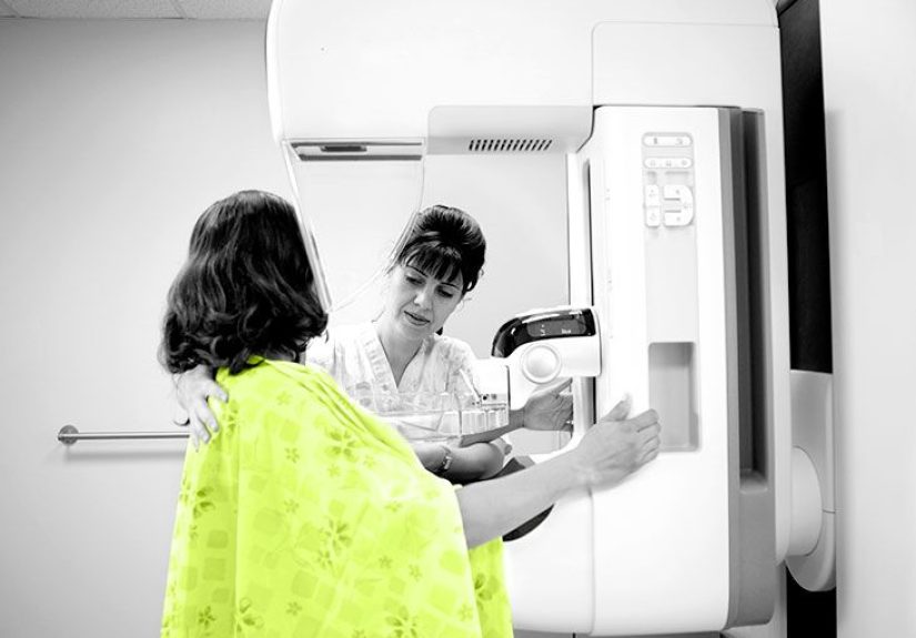
Mammogram: looking for patterns, calcifications, and hidden issues
Mammography uses low-dose X-rays to evaluate breast tissue. It can detect masses, asymmetries, and calcifications,
and it’s often paired with ultrasound for a fuller pictureespecially when a lump is palpable.
Aspiration: “If it’s fluid, we can prove it”
If imaging suggests a cyst (or if a lump is painful and likely cystic), a clinician may perform a fine-needle aspiration.
If fluid comes out and the lump collapses, that’s strong evidence it was a cyst. Sometimes the fluid is sent to the lab,
especially if it’s bloody or the imaging features aren’t straightforward.
Biopsy: the final answer when cancer can’t be ruled out
When imaging looks suspiciousor when a solid mass needs confirmationclinicians may recommend a core needle biopsy
(or another biopsy approach). A biopsy is the way to determine whether cells are benign, atypical, or cancerous.
Translation: imaging can be highly informative, but tissue diagnosis is what confirms cancer.
Red flags: when to call a clinician sooner rather than later
Any new breast lump should be evaluated, but these signs deserve prompt medical attention:
- Skin dimpling or puckering
- Orange-peel texture (peau d’orange)
- Nipple retraction (pulling inward) that’s new
- Bloody nipple discharge
- Persistent, enlarging lump over weeks
- Swelling of part or all of the breast
- Enlarged lymph nodes in the armpit or near the collarbone
- Redness, warmth, fever (could be infectionstill needs evaluation)
What happens after you get answers
If it’s a simple cyst
- No treatment if it’s not bothering you.
- Aspiration if it’s painful or large.
- Follow-up if it recurs often or has atypical features.
If it’s complicated or complex
Your clinician may recommend short-interval imaging follow-up, aspiration, or biopsydepending on what the ultrasound shows
and your overall risk factors.
If it’s a benign solid tumor (like fibroadenoma)
Many are monitored with periodic imaging to confirm stability. Removal might be discussed if it grows, causes symptoms,
or has features that make the diagnosis less certain.
If it’s cancer
The next steps typically involve staging workup and a treatment plan that may include surgery, radiation, hormone therapy,
targeted therapy, chemotherapy, or a combinationtailored to the cancer type and stage.
Quick FAQ (because your brain will ask these at 2 a.m.)
“Can a breast cyst turn into cancer?”
A simple cyst itself is generally considered benign. The bigger clinical issue is whether a complex-looking “cystic” mass
actually contains solid components that need evaluation. That’s why ultrasound descriptors matter so much.
“If it hurts, is it more likely a cyst?”
Pain can happen with cysts and fibrocystic changes, but pain alone doesn’t prove it’s a cyst or rule out cancer.
Persistent focal pain should still be evaluated.
“If it moves, is it definitely not cancer?”
Not definitely. Mobility can suggest a benign process, but it isn’t a guarantee. Imaging decides; biopsy confirms when needed.
“What should I say at my appointment?”
Bring specifics: when you noticed it, whether it changes with your cycle, whether there’s pain, nipple discharge,
skin changes, recent pregnancy/breastfeeding, family history, and any prior breast imaging or biopsies.
Real-world experiences: what this often feels like (the human side)
Let’s talk about the part nobody puts on the lab report: the waiting. People often describe a new breast lump as an instant anxiety amplifier.
Even if you’re usually calm, a lump can make your mind sprint to worst-case scenariosbecause the stakes feel personal and immediate.
And honestly? That reaction is normal.
One common experience with cysts is the “it showed up out of nowhere” moment. Someone is showering or changing clothes,
feels a round, tender lump, and suddenly the day is divided into “before lump” and “after lump.” At the clinic, an ultrasound may show
a classic fluid-filled pocket. If the cyst is large or painful, aspiration can be offered. Many people describe the relief as almost comical:
the lump that felt like a major life event turns out to be fluid, and it literally disappears in minutes. The emotional whiplash is real.
Another frequent story: a solid lump that feels smooth and movableoften a fibroadenomafollowed by the world’s least fun encore:
“Let’s recheck it in 6 months.” Monitoring can be medically appropriate, but emotionally, it can feel like being told,
“Good news! Nothing is on fire. We will now schedule periodic fire checks.” People cope by making the follow-up plan concrete:
put the imaging date on the calendar, write down questions, and ask the radiology team what changes should trigger an earlier call.
For those whose imaging results are unclear, the biopsy experience is often described as more intimidating in theory than in reality.
Many patients report that the scariest part is the word biopsy, not the procedure itself. The actual processlocal anesthesia,
a little pressure, some clicking soundstends to be manageable. What’s harder is waiting for pathology results.
A practical tip people share: schedule something gentle but absorbing for the day after (a walk with a friend, a comfort-show marathon,
an anything-but-Google activity).
People who’ve been through this also tend to become unexpectedly good at advocating for themselves.
They learn to ask questions like: “Is this fluid-filled or solid?” “What category is it?” “Do I need follow-up imaging?”
“If we’re watching it, what changes should I report?” That shiftfrom fear to a planoften helps more than any single reassurance.
Finally, there’s a quiet but important theme: most breast lumps are not cancer, but every lump deserves respect.
The goal isn’t to self-diagnose; it’s to get the right evaluation efficiently, so you can move from uncertainty to clarity.
If you’re reading this with one hand on your phone and the other doing an anxious “lump re-check,” consider this your sign to book the appointment.
Your future self will thank you.
Conclusion
If you remember only three things: (1) cysts are usually fluid-filled and often benign, (2) tumors are solid and can be benign or malignant,
and (3) ultrasound and, when needed, biopsy are how you truly tell the difference.
Your job isn’t to guess correctlyit’s to show up with good notes, get the right imaging, and follow the plan.