Table of Contents >> Show >> Hide
Note: This article is for educational purposes only and is not a substitute for professional medical care. If someone has vomiting of blood, black tarry stools, severe abdominal swelling, confusion, or trouble breathing, that is not the moment for a search engine deep dive. That is the moment to get urgent medical help.
Caput medusae sounds like the title of a lost mythology sequel, but in medicine it describes a very real and very important physical sign: enlarged veins spreading across the abdomen, usually around the belly button, in a pattern that can resemble the snake-covered head of Medusa. It may look dramatic, but the bigger issue is what it often points to behind the scenes. In many cases, caput medusae is a clue that pressure has built up in the portal venous system, the network of veins that carries blood from the digestive organs to the liver.
That means caput medusae is not a disease by itself. It is a warning light on the dashboard. And like every warning light, it matters most because of what may be happening underneath it: cirrhosis, portal hypertension, blocked blood flow, or other serious problems involving the liver and major abdominal veins. In this article, we will break down what caput medusae is, what symptoms often show up with it, what causes it, how doctors diagnose it, and what treatment usually focuses on. We will also add practical, real-world context so the topic feels less like a textbook and more like something an actual human can understand before their coffee gets cold.
What Is Caput Medusae?
Caput medusae is the visible appearance of swollen, enlarged veins on the surface of the abdomen, especially around the navel. These veins are collateral veins, meaning they act like backup routes when blood cannot move normally through its usual pathway. Think of it as traffic being rerouted off a blocked highway and onto side streets. The side streets were not built for that much traffic, so they swell, twist, and become more obvious.
The most common reason this happens is portal hypertension, which means increased pressure in the portal vein system. The portal vein carries blood from the stomach, intestines, pancreas, spleen, and related organs to the liver. When scarring, clotting, or another blockage slows this blood flow, pressure rises. The body then opens alternate pathways, including paraumbilical veins near the belly button, and those veins can become prominent enough to create the classic caput medusae pattern.
So, no, your abdomen is not trying to audition for Greek mythology. But it is sending a message that deserves medical attention.
Symptoms of Caput Medusae
What it looks like
The main visible sign is a network of bluish, swollen, tortuous veins spreading outward from the belly button or across the front of the abdomen. Some people notice them all at once in the mirror. Others first feel that something “looks off” during bathing, dressing, or changing clothes. The veins themselves are often painless, which can make the sign seem oddly harmless at first glance.
What it may feel like
Caput medusae itself usually does not cause severe pain. But the condition causing it often does bring other symptoms. Those can include:
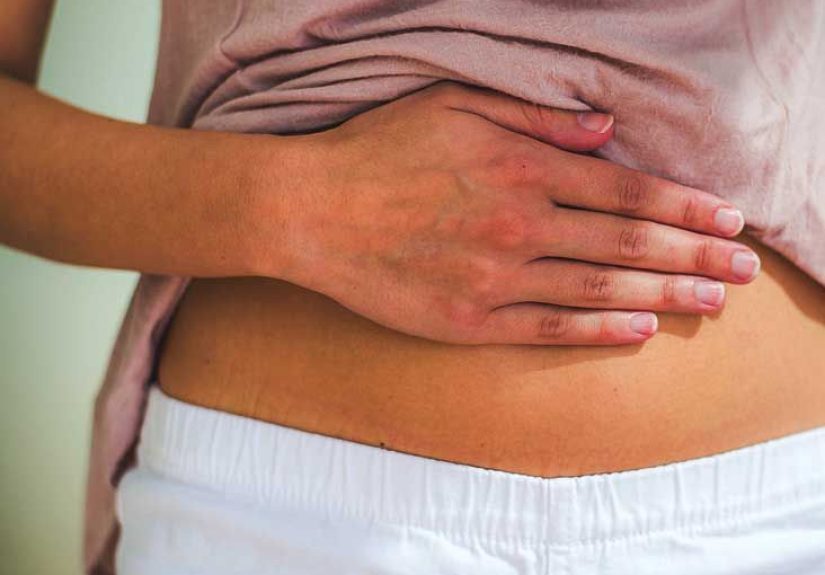
- Abdominal swelling from ascites, which is fluid buildup in the abdomen
- Swelling in the legs, ankles, or feet
- A feeling of fullness or pressure in the belly
- Abdominal discomfort or pain
- Fatigue and weakness
- Yellowing of the skin or eyes, also known as jaundice
- Itchy skin, dark urine, or pale stools in advanced liver disease
- Confusion, sleep changes, or “brain fog” related to hepatic encephalopathy
- Vomiting blood or passing black, tarry stools if varices bleed
In many patients, the swollen abdominal veins are not the first symptom of liver disease. They arrive later, when portal hypertension has become more significant. That is why caput medusae often signals a problem that is already more than mild.
What Causes Caput Medusae?
The most common cause: portal hypertension
Most cases of caput medusae are linked to portal hypertension, especially when that pressure increase comes from cirrhosis. Cirrhosis is severe scarring of the liver. Scar tissue makes it harder for blood to flow through the liver normally, so pressure backs up in the portal vein system. When that happens, blood finds alternate routes, including veins near the umbilicus.
In the United States, common reasons people develop cirrhosis include chronic hepatitis B or hepatitis C, alcohol-related liver disease, and metabolic dysfunction-associated steatotic liver disease, the condition previously known under the NAFLD umbrella. Over time, these conditions can damage the liver enough to create the pressure problem that leads to caput medusae.
Other possible causes
Although portal hypertension from cirrhosis is the usual suspect, it is not the only one in the lineup. Other causes can include:
- Portal vein thrombosis, meaning a clot blocks blood flow in the portal vein
- Budd-Chiari syndrome, which involves blocked hepatic venous outflow
- Inferior vena cava obstruction, which can also redirect blood through abdominal veins
- Less common forms of noncirrhotic portal hypertension
- Certain infiltrative, inflammatory, or vascular liver conditions
This is why doctors do not stop at saying, “Yep, those are enlarged veins.” The real work is figuring out why they are there.
How Caput Medusae Is Diagnosed
1. History and physical exam
Diagnosis often begins with a careful medical history and a physical exam. A clinician will look at the abdomen, check for visible venous patterns, and ask about liver disease risk factors such as heavy alcohol use, viral hepatitis, prior clotting problems, unexplained swelling, weight changes, or past episodes of jaundice. They may also look for an enlarged liver, enlarged spleen, leg edema, or ascites.
2. Blood tests
Blood work does not diagnose caput medusae directly, but it helps uncover the cause. Common tests may include liver enzymes, bilirubin, albumin, clotting studies, complete blood count, kidney function, and tests for viral hepatitis or iron overload. These results help reveal whether liver injury, impaired liver function, or another systemic issue is part of the picture.
3. Imaging studies
Doppler ultrasound is often the first imaging test used because it can show blood flow in the portal vein and surrounding vessels without being invasive. It can also help distinguish portal hypertension from other types of venous obstruction by showing the direction of blood flow.
Doctors may also order:
- CT scan of the abdomen
- MRI or MR venography
- Ultrasound to look for ascites, splenomegaly, or liver texture changes
These studies can show collateral vessels, clots, cirrhotic changes, and other structural clues.
4. Endoscopy and pressure studies
If portal hypertension is suspected, an upper endoscopy may be done to check for esophageal or gastric varices. This matters because those veins can bleed heavily and sometimes without much warning.
In select cases, specialists may measure the hepatic venous pressure gradient to assess how high the portal pressure is. This is not required for every patient, but it can help in complex or specialized cases.
Treatment for Caput Medusae
Treat the cause, not just the veins
Caput medusae itself usually does not need cosmetic treatment. The goal is to treat the condition behind it. In other words, the veins are the messenger, and medicine is interested in the message.
Managing portal hypertension and cirrhosis
Treatment depends on what is driving the portal hypertension and whether complications have developed. Common approaches include:
- Nonselective beta blockers such as propranolol or nadolol to reduce portal pressure and lower the risk of variceal bleeding
- Sodium restriction and diuretics to help control ascites and leg swelling
- Paracentesis to drain large-volume ascites when the abdomen becomes tense or uncomfortable
- Lactulose and sometimes rifaximin for hepatic encephalopathy
- Treatment of the underlying liver disease, such as antivirals, alcohol cessation support, or metabolic risk reduction
When bleeding risk is high
If a patient has esophageal varices, doctors may recommend endoscopic band ligation. This procedure places small rubber bands on enlarged veins in the esophagus to reduce bleeding risk. If there is active bleeding, emergency treatment may include IV medications such as octreotide, blood transfusions, endoscopy, and hospital-level care.
Advanced procedures
If portal hypertension remains severe or complications keep recurring, a doctor may recommend a TIPS procedure, short for transjugular intrahepatic portosystemic shunt. In this procedure, a radiologist creates a channel inside the liver to divert blood flow and reduce pressure in the portal system. TIPS can be very effective, but it also has risks, including worsening encephalopathy in some patients.
In advanced liver disease, especially when cirrhosis is decompensated, liver transplantation may become the definitive treatment option.
When to Seek Urgent Medical Care
Caput medusae should not be ignored, but some related symptoms are especially urgent. Seek immediate medical care if there is:
- Vomiting blood
- Black or bloody stools
- Sudden severe abdominal swelling or pain
- Confusion, extreme sleepiness, or new disorientation
- Shortness of breath
- Marked decrease in urine output
- Rapid worsening of jaundice
Those symptoms can signal bleeding varices, infection, worsening liver failure, kidney complications, or severe portal hypertension. In plain English: not a “wait and see” situation.
Can Caput Medusae Be Prevented?
Prevention is really about preventing the underlying disease from progressing. That can mean getting treated for hepatitis, reducing or stopping alcohol use, managing obesity and metabolic risk factors, following up on abnormal liver tests, and staying engaged with routine medical care if cirrhosis is already present. Earlier treatment of liver disease can sometimes prevent the chain reaction that leads to portal hypertension and visible collateral veins.
Real-World Experiences Related to Caput Medusae
In real life, the experience of caput medusae is often less dramatic at first than the name suggests. Many people do not walk into a clinic saying, “I think I have portal hypertension.” They say something more like, “I noticed weird veins around my belly button,” or “My stomach looks different, and I’m not sure why.” Sometimes a family member spots it first. Sometimes it shows up gradually enough that the person normalizes it until another symptom, like swelling, fatigue, or jaundice, forces the issue.
For patients with undiagnosed liver disease, seeing those veins can be unsettling. The common reaction is a mix of curiosity and denial. They may search online, hope it is “just varicose veins,” and then discover that caput medusae is usually linked to bigger circulation or liver problems. That moment can be scary, but it is also often the moment they finally seek evaluation. In that sense, the visible veins become an important clue that leads to diagnosis.
For people already living with cirrhosis, caput medusae can feel like one more visible reminder that the disease is no longer abstract. It is no longer just a lab result, a scan report, or a conversation with a hepatologist. It is something visible on the body every day. Some people describe feeling embarrassed at the beach, self-conscious in front of a partner, or anxious every time they notice the veins looking more prominent. Others say the appearance itself does not bother them much, but the associated symptoms do: the tightness of ascites, the exhaustion, the constant need for follow-up appointments, and the fear of bleeding.
Clinicians often see a pattern in these stories. A patient comes in because of abdominal swelling, swollen legs, confusion, poor appetite, or a bleed, and during the exam the provider notices caput medusae. The finding helps connect the dots. It tells the team that portal pressure may be high and that the patient should be evaluated for complications such as varices, ascites, or worsening liver function. In that setting, the veins are not just a curiosity. They are a clue that changes how urgently the rest of the workup needs to happen.
There is also a practical side to the experience. Once treatment starts, patients often realize that managing the underlying condition is a long game. They may need medication changes, endoscopy appointments, sodium restriction, alcohol cessation counseling, monitoring for liver cancer, or transplant evaluation. The visible veins may not disappear quickly, and sometimes they do not go away completely. That can be frustrating if someone hoped treatment would make everything look normal right away. But from a medical standpoint, success is measured less by cosmetic change and more by improved portal pressure, fewer complications, and better liver stability.
What many patients and families say helps most is having the problem explained in clear language. When they understand that caput medusae is a sign of rerouted blood flow, not a random surface issue, the whole situation becomes easier to grasp. And once the “why” becomes clearer, the next steps feel less mysterious. The experience is still serious, but it becomes manageable in a way that raw fear never is.
Final Takeaway
Caput medusae is one of those medical signs that looks striking because it is striking. Enlarged abdominal wall veins are usually a sign that blood is being rerouted because normal flow through the liver or major abdominal veins is obstructed. Most often, that means portal hypertension, frequently due to cirrhosis. Diagnosis usually involves physical examination, blood tests, Doppler ultrasound, and often imaging or endoscopy. Treatment focuses on the underlying cause and on preventing dangerous complications such as variceal bleeding, ascites, encephalopathy, and liver failure.
The key point is simple: caput medusae is not something to shrug off. It is a visible sign that something significant may be happening inside the abdomen. When seen early and evaluated properly, it can help doctors identify serious liver or vascular disease before complications become even more dangerous.